

SELEX Galileo Scientific
Photo Competition 2010
Organized by School of Engineering and Physical Sciences
•
Sponsored by SELEX Galileo
and School of EPS, Heriot-Watt
University
•
145 submitted photos and 38 participants: all the submitted photos
can be found here
Title: Quantum
State Engineering and Detection
Adetunmise Dada
Cavity quantum electrodynamics (QED) implementation
of a joint generalized quantum measurement of two atoms. This illustration shows
two two-level atoms flying through high-Q cavities and Ramsey zones, bouncing
off magnetic mirrors, and finally passing through field ionizing
detectors. This is a promising technique for quantum
information technologies.

Title: Testtubes
Ai-lan Lee
Rack of test tubes in a typical chemistry lab
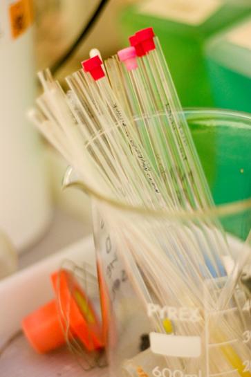
Title: Chemistry Lab
Ai-lan Lee
NMR sample tubes in a typical chemistry lab

Title: Periodic Table
Ai-lan Lee
Periodic Table
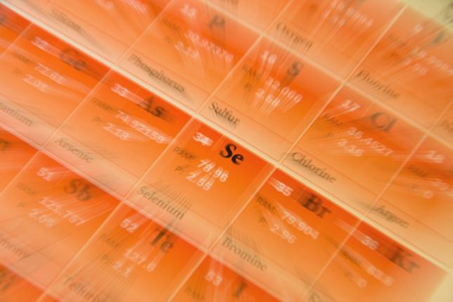
Title: Selenium Burst
Ai-lan Lee
Zoom burst of the element selenium in the Periodic Table
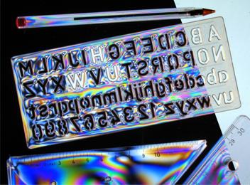
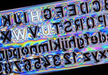
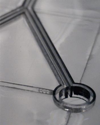
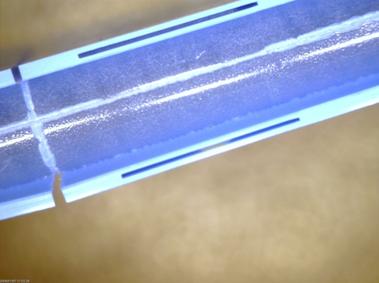
Title: Made to measure stencil: HWU
shines through!
Aongus McCarthy
School of EPS
Viewing transparent moulded plastic items between
crossed polarisers can reveal colourful stress patterns because of the induced
birefringence. The symmetry of the H-W-U letters in this plastic stencil meant
that they looked the same, and were in the “right” order, when the stencil was
turned over! The letters were made to appear bright between crossed polarisers by
using pieces of sellotape which cause the plane of polarisation to be rotated.
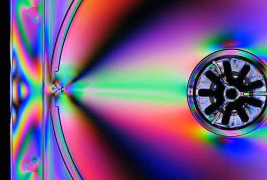
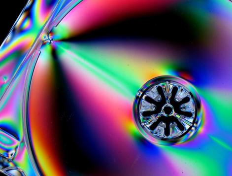
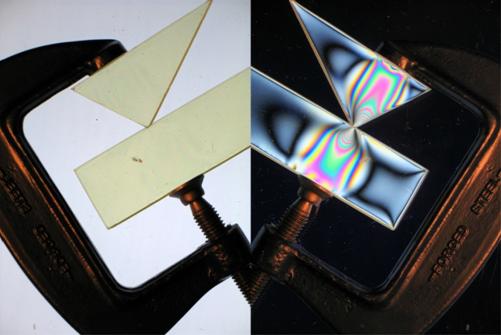
Title: A compact stress recording
Aongus McCarthy
School of EPS
Viewing transparent moulded plastic items between
crossed polarisers can reveal colourful stress patterns because of the induced
birefringence. This was first described by the Scottish physicist Sir David
Brewster in the early 19th century. These photographs shown a CD jewel case
insert tray back illuminated with polarised light and photographed through a polarising
filter attached to the camera.
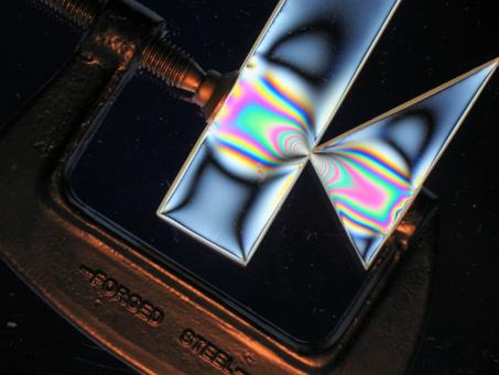
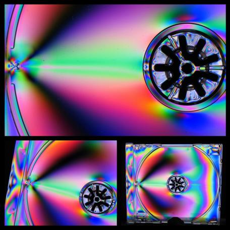
Title: Clamp down on stress
Aongus McCarthy
School of EPS
Certain transparent materials reveal colourful
contour patterns when they are placed under mechanical stress and viewed
between crossed polarisers. The patterns are visible because of the induced
birefringence which is proportional to the applied stress. This is known as
mechanical birefringence or photo elasticity, and was first described by the
Scottish physicist Sir David Brewster in the early 19th century.
Title: Optical highways - No traffic lights required at the intersections
Aongus McCarthy
School of EPS
There is considerable interest in incorporating
"optical pathways", within metre-scale; electrical printed circuit boards
(PCBs). This photograph shows optical waveguides (50 μm square cross-section
cores with cladding) that have been formed in polymer on a PCB substrate
material using a UV laser-writing system. (Scale: The radius of curvature of
the smaller bends is approximately 15 mm).
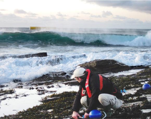
Title: EMEC wave test site
Beharie, Robert A
The photo was taken by a colleague of
the author at the EMEC wave test site. Whereas he processed the image to its
final version making it a collaborative process. The subject is research into
wave energy by using innovative techniques. In the background is the Aquamarine
wave energy converter device.
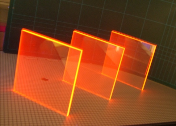
Title: Novel Europium complex with
record quantum efficiency in polymer matrix
Brenda C Rowan
A novel
fluorescent Europium complex with record efficiency in a polymer matrix (86%)
is shown. Under UV illumination the bright edge emission due to total internal
reflection can be seen. Applications being investigated include luminescent
solar concentrators, spectral shifting to improve solar cell efficiency and
improvement of algal growth conditions.

Title: Electrical Droplet running Track
R FU
Electrical Driven
patterns have been generated on Si substrate, which can drive liquid droplet
running within the track.
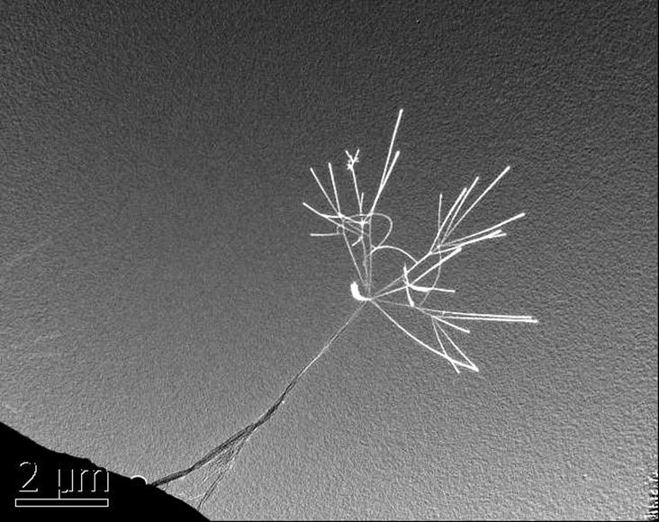
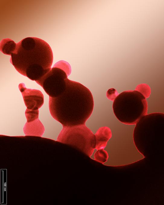
Cabon Nanotube flower
R Fu
Transmission electron
microscopy image showing the field Emission of Carbon naotube. When the
electron beam shines on the carbon nanotube, the field emission causes the
“flowering” charging effect.
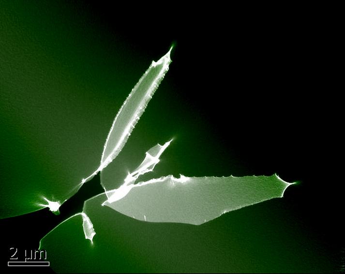
Cabon Nanotube Sword
R Fu
Transmission electron
microscopy image showing the field Emission of Carbon naotube. When the
electron beam shines on the carbon nanotube, the field emission causes the
charging effect.
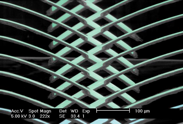
Rowing of microcantilevers
Richard Fu
Nano Disney world
R Fu
Transmission electron
microscopy image of nano-silver particle shows the self-sintering and necking
of nanoparticle surface due to size effect. In his image, the nanoparticles
form patterns similar to Disney characters.

Nano-dog
TEM images of
nanoparticles
By Richard FU


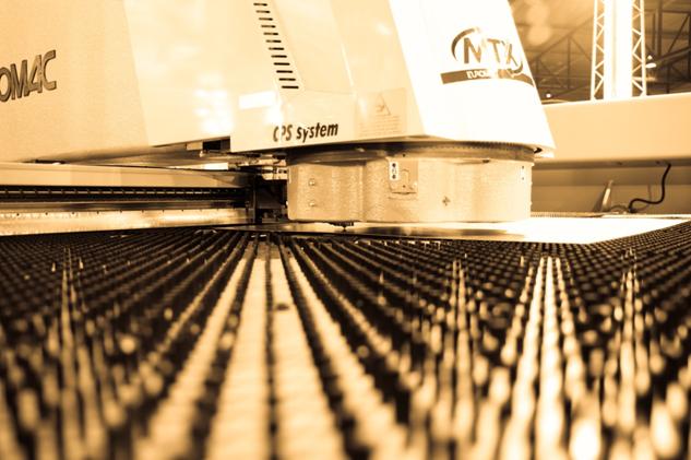
Cmihovs Aleksejs
The press machine for cutting out
different shapes from steel plate.

Cmihovs Aleksejs
A photo of Engineers best friend
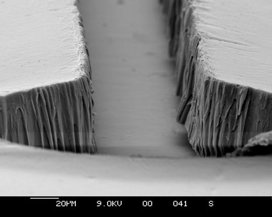
Microchannels
for Blood Cell Separation.
Deirdre Kavanagh
This image shows an unpackaged 100µm
microfluidic channel with circular reservoirs, fabricated in the negative
photoresist SU8. SU8 is spincoated on a 200nm titanium seed layer on top of a
glass wafer and patterned using UV lithography. The channel will be implemented
in a lab on chip for non-invasive prenatal diagnosis. Photo has
been cropped.
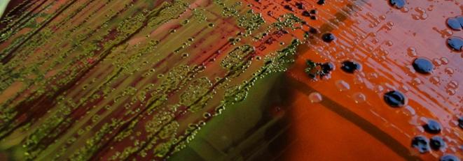
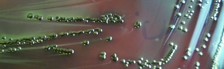
Title:
Metallica
Dewar
Susan J
The presence of the
potential foodborne pathogen E. coli
on these chromogenic L-EMB plates is shown by the distinctive metallic sheen of
the circular colonies on the agar surface.
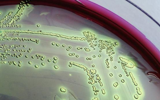
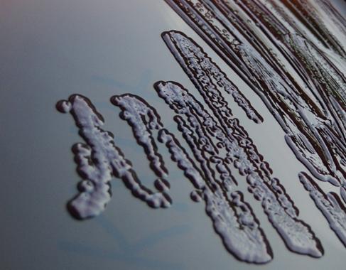
Title:
Rise of the Clones
Dewar
Susan J
The round, glutinous
bacterial colonies that can be seen on the surface of this microbiological plate
arise from single progenitor cells and comprise clonal populations of the
organism being isolated.
Title:
Bacterial Tartan
Dewar
Susan J
The environment around us
contains a diverse microflora that exists largely without our noticing. This
plates shows just a few of the many, many bacterial species that add interest
and a touch of colour, although usually hidden, to our lives.
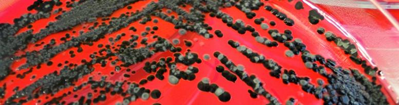
Title:
Liquorice Allsorts
Dewar
Susan J
Advances in culture media
formulation allow the rapid isolation and detection of particular pathogens.
Chromogenic agars like the one shown here contain substrates that are broken
down by bacteria to allow the production of all sorts of species-specific
colours, like the black of the Salmonella colonies shown here.
Title:
Bad Press
Dewar
Susan J
The majority of E. coli
bacteria we come into intimate contact with are harmless commensal organisms
that help support our digestion. This photo shows microbiological plates confirming
the presence of E. coli, an organism that gets a lot of bad press that’s
justified by only a small number of its extended family.
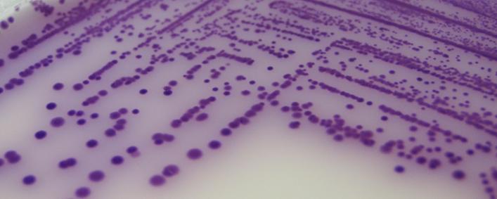
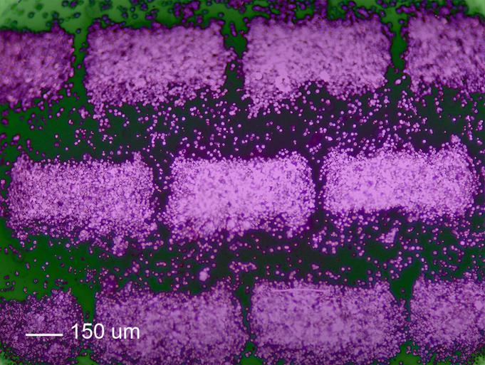
Title:
Purple Rain
Dewar
Susan J
The presence of any number
of Salmonella cells in a food makes it unacceptable for consumption.
BrillianceTM Salmonella Agar incorporates a novel inhibitor technology that
allows the isolation and presumptive identification of Salmonella species, as
evidenced by the distinctive purple colour of the Salmonella colonies seen in
the photo.
Title: Digital Micro-fluidic
Biosensor
Douglas Stuart Brodie
Integrated
platform using Surface Acoustic Wave technology and Electro Wetting on
Dielectric (EWOD) to transport and mix fluid before being moved onto a reaction
platform where sensitive bio-sensing measurements can be made.
Title: Digital Micro-fluidic
Biosensor
Douglas Stuart Brodie
Integrated
platform using Surface Acoustic Wave technology and Electro Wetting on
Dielectric (EWOD) to transport and mix fluid before being moved onto a reaction
platform where sensitive bio-sensing measurements can be made.
Title: Surface Acoustic Waver
Tweezers
Douglas Stuart Brodie
Heriot-Watt design for acoustic tweezers which can immobilize biological
material within a solution into a predefined pattern formed by the acoustic
wave. Shown in the example is blood cells
Title: Surface Acoustic Waver
Tweezers
Douglas Stuart Brodie
Heriot-Watt design for acoustic tweezers which can immobilize biological
material within a solution into a predefined pattern formed by the acoustic
wave. Shown in the example is blood cells
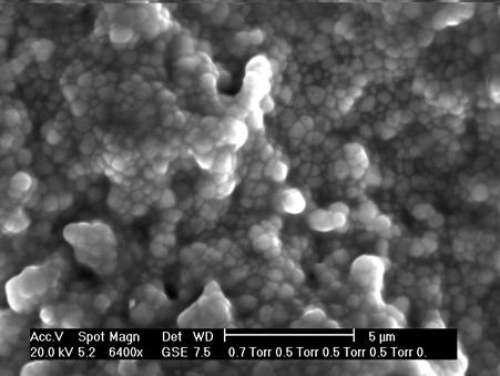
Title:
ESEM
(Environmental Scanning Electron
Microscope) image of the rough surface 3Y-TZP
(Ytria-Stabilized Tetragonal Zirconia Polycrystal) ceramics.
Title: Light scattering
of ceramics
Light
scattered by multicrystalline structure in 3Y-TZP (Ytria-Stabilized Tetragonal
Zirconia Polycrystal) block.
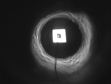
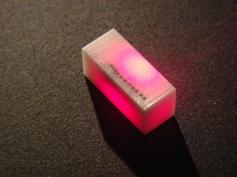
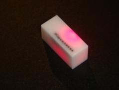
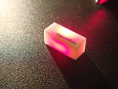
Title: LED emitter
Light
emission from the LED emitter observed using macro lens.
The square emitter is surrounded by the standard 3 mm epoxy
dome lens. The wire connects the anode (+) with the emitter mounted
on the cathode.
Artworks submitted by: Mateusz Matysiak
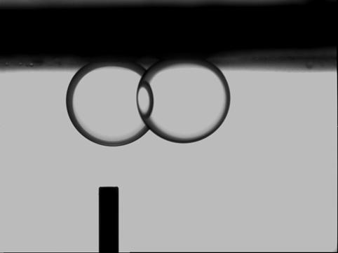
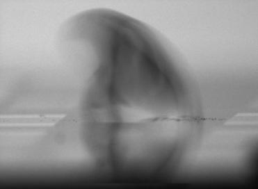
Title: Interaction of fluid-fluid and fluid-solid at their
interfaces
Interfacial
forces and wettability of the solid-oil-water-carbon dioxide system play an
important role in a number of environmental, biological and industrial
processes. Measuring the aforementioned data under high pressure-high
temperature conditions is essential to design enhanced oil recovery and CO2
storage processes. This photo was taken during one of our experiments, which
shows almost two same sized oil droplets with different distance from the
camera and light source. The common area is a guidance to clarify the order of
each droplet in terms of position from light source.
Title: Interaction of
fluid-fluid and fluid-solid at their interfaces
Interfacial
forces and wettability of the solid-oil-water-carbon dioxide system play an
important role in a number of environmental, biological and industrial
processes. Measuring the aforementioned data under high pressure-high
temperature conditions is essential to design enhanced oil recovery and CO2
storage processes.
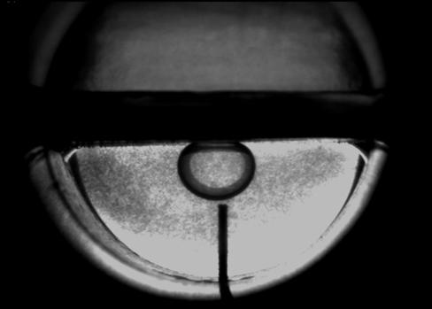
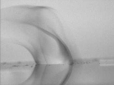
Title:
Micro gas bubbles in water
Interfacial
forces and wettability of the solid-oil-water-carbon dioxide system play an
important role in a number of environmental, biological and industrial
processes. This picture shows micro-gas bubbles inside water as a result of
pressure drop of the system. As can be seen due to very small size of the gas
bubbles light can not pass through and case a shadow on the image of the media,
which is clear water.

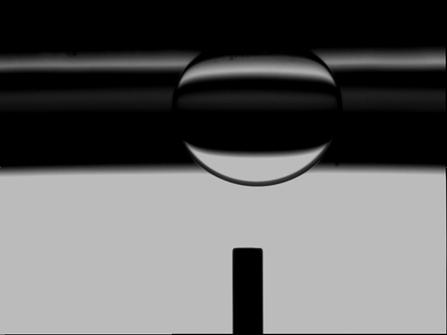
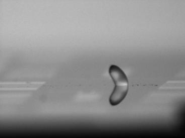
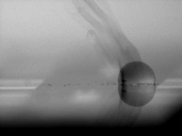
Title:
Partial eclipse in the lab
Interfacial
forces and wettability of the solid-oil-water-carbon dioxide system play an
important role in a number of environmental, biological and industrial
processes. In one of the experiments we tested the penetration of light from
fluids and observed that due to the special shape of the oil droplet it can
pass the light and generate a speculate picture as if it is the picture of moon
and/or a real partial eclipse of sun.
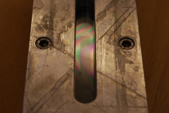
Title:
Transparent porous medium
This
rainbow is due to the reflection of light from one of our equipment which is
made of two pieces of glass. The different thicknesses of tapped air between
these two glass plates are the main reason for this reflection.
Artworks submitted by: Masoud Riazi
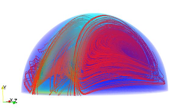
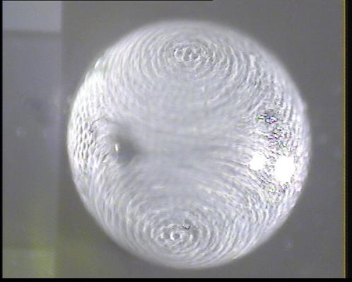
Title: experiment and
simulation for surface acoustic wave Microfluidic device
Experimental
picture from top view for surface acoustic wave impinging liquid droplet from
right hand side which results in butter-flay streaming patterns. 3D numerical
simulation results for SAW Microdroplet interaction. In this pictures
butter-flay streaming patterns are presented.
Artworks submitted by:
Mansuor Alghane
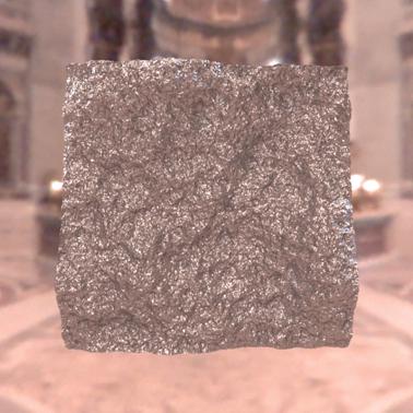
Title: A computer synthetic visual image
A
glossy surface is rendered using real world image lighting, showing a glossy
appearance. This provides us a powerful tool to study people’s perception of these
visual effects.
Artworks submitted by:
Lin Qi
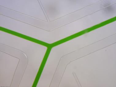
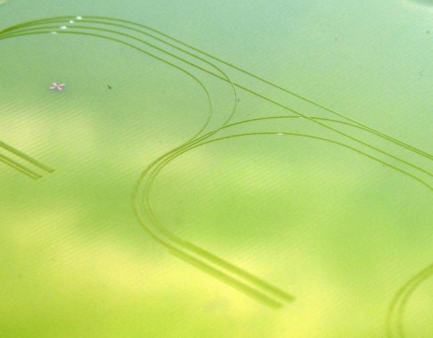
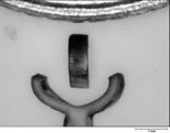
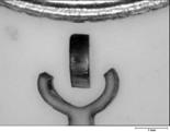
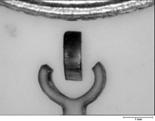
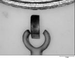
Title:
A perfect day
This
photograph shows a Y-shaped PDMS (polydimethylsiloxane, a type of polymer )
Microfluidic channel filled with green dye. The channel is 100 microns wide and
leak-free. The channel walls are extremely smooth. The green colour and the
shape of the channel reminding of the peace symbol, make me think that it was
just “a perfect day”.
Artworks submitted by:
Kersaudy Kerhoas Maiwenn
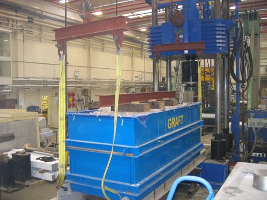
Title:
GRAFT facility being moved
GeoPavement
& Railway Accelerated Fatigue Testing (GRAFT) facility being positioned by
overhead cranes underneath a cyclic loading actuator. GRAFT is a unique
facility that has been developed over the last three years to test the
performance of railway trackbeds under loading. The results have direct
implications for reducing track maintenance.
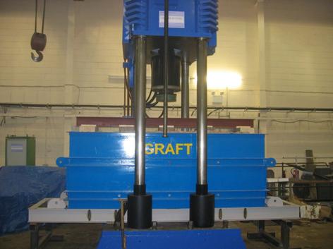
Title: GRAFT facility
ready to be loaded
GeoPavement
& Railway Accelerated Fatigue Testing (GRAFT) ready to be loaded from a 200
tonne cyclic actuator. GRAFT is a unique facility that has been developed over
the last three years to test the performance of railway trackbeds under
loading. The results have direct implications for reducing track maintenance.
Artworks submitted by:
Justin Kennedy
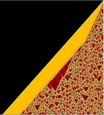
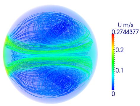
Title: Invasion dynamics in an oscillatory system
In
oscillatory systems, invasion generates a spatiotemporal transition through
periodic cycles,leading ultimately to spatiotemporal chaos.
Title:
Waves and Chaos in Oscillatory Systems
Many
natural populations undergo multi-year cycles, and field studies have shown
that these
can be organized into periodic travelling waves. Mathematical studies have
shown that large-scale landscape obstacles represent a natural mechanism for
wave generation. This figure show a numerical simulation of this process of
wave generation for a caricature model of an oscillatory ecological system. In
the left hand panel the obstacle is the small central circle, and it generates
a stable wave pattern. But in the right hand panel the obstacle is larger, and
this causes the generation of a wave that is of lower amplitude and is
unstable. It is visible close to the obstacle but then breaks down into
spatiotemporal chaos. In oscillatory systems, invasion generates a spatiotemporal
transition through periodic cycles,leading ultimately to spatiotemporal chaos.
Artworks submitted by: Jonathan Sherratt and Matthew Smith
Title: Hologram produced by a Spatial Light Modulator
The phase
of the laser light has been selectively modified by a Spatial Light Modulator
to produce an image at the focus of an optical system. The light can be
seen to revert back to a spatial frequency distribution in the far distance.
Artworks submitted by: Jonathan Parry
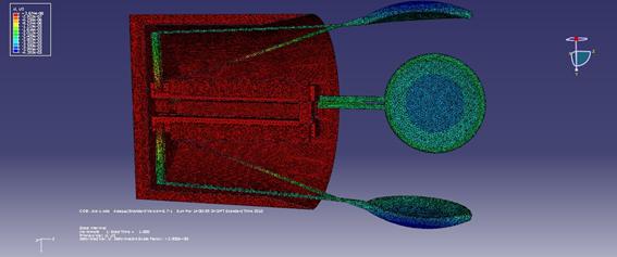
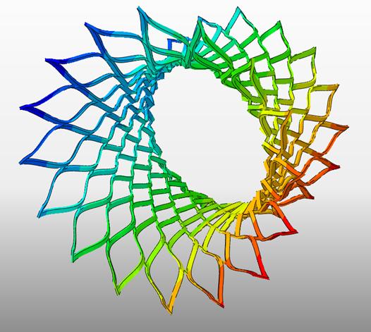
Gripper
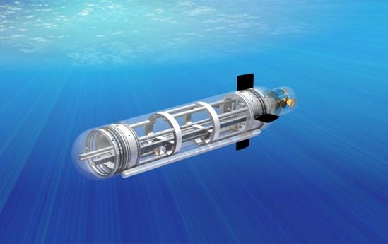
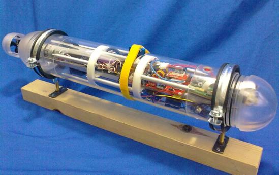
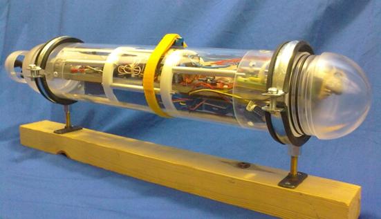
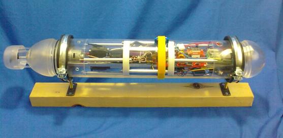
The images show forth year
project, mAUVe, which stands for micro Autonomous Underwater Vehicle for
exploration, it measures about 750mm long for scale. There is also a the cad
models for this project along with some FEA analysis for a 3D micro Gripper
which has been an interesting learning experience giving me a much better
understanding of FEA and abaqus.
Artworks submitted by: Jonathan Clay
Title: Expanded Coronary Stent
A
Finite Element Analysis of a Coronary Stent, which is used in heart surgery to
open partially blocked arteries. The picture shows an analytical
deformation of the stent, which is vital, along with stress analysis, in the
design process.
Artworks submitted by: James Young

Title:
The demonstration of remote micro-clamp by thermal triggering
The
demonstration show the potential application on micro-device manufacture based
on current shape memory materials, those PICs provided the visual evidences
about the shape memory properties and also the potential application based on
this smart materials.
Artworks submitted by: Bin Xu
Title:
The jumping of micro-droplets
The
micro droplets jumped with the outer applied electrical field, series pics show
the movement of droplets
Title:
The micro mixing in single micro droplets
The
mixing procedure in this pic reveal a important micro fluid behaviour.
Artworks submitted by: Yifan Li
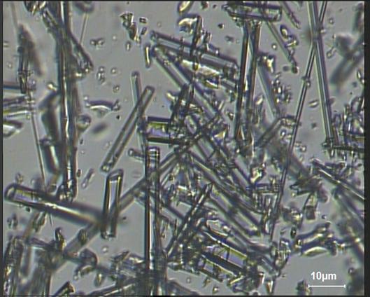
Title: Fabrication of Microneedles for
Microinjection
Muriel
Béchu
School of EPS
Glass
capillaries were heated and drawn in a micropipette puller and the thinnest
ends of the resulting needles cut with a razor blade. The average diameter of a
microneedle is about 3µm. These microneedles can be optically tweezed and might
some day be an alternative to mechanical microinjection into cells.
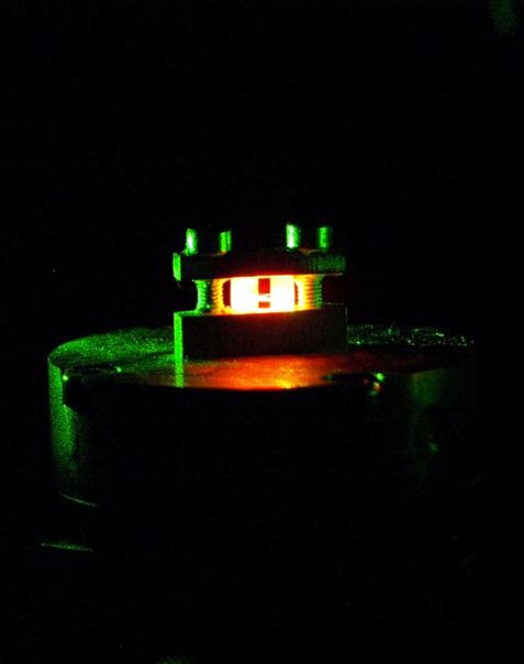
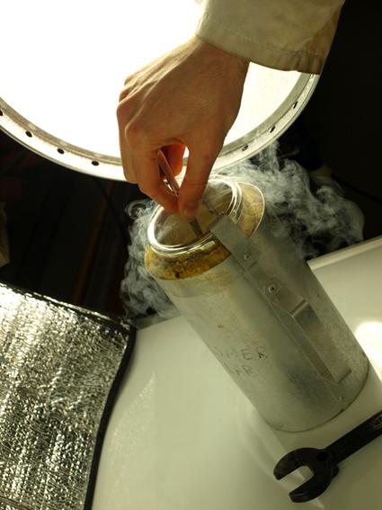
Title: High speed optics lasers for
metrology
Nikolaj
Rybakov
School of EPS
The
photo with black background where 2 bolts are holding a glowing red crystal is
the same laser setup, the crystal is reflecting a powerful laser coming out
directly from the generator. The interesting thing that the laser wasn’t picked
up by the camera however something different was. And the photo with a hand is
taken from chemistry department, the guy is cooling a rubber bit in -172C in
liquid nitrogen.
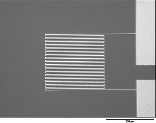
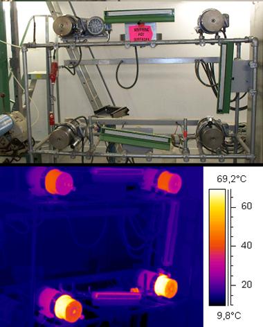
Title: Micro Temperature Sensor with
Footprint of 250µm×240µm
Yufei
Liu
School of EPS
For
in-situ temperature monitoring of laser assisted bonding for MEMS packaging
application, thin film micro sensors have been designed with meander track
width of 3µm resulting in a footprint of 250µm×240µm, which were fabricated by
using sputtered platinum and patterned using the focused ion beam (FIB) etching
method.
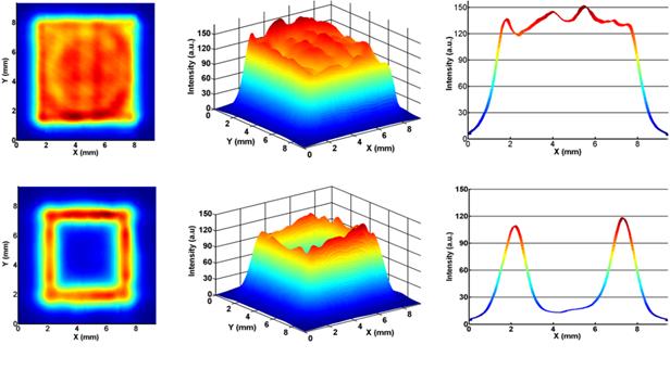
Title: Beam Profiles of Laser Assisted
Bonding for MEMS Packaging
Yufei
Liu
School of EPS
Localised
laser assisted bonding can reduce the thermal load on MEMS devices and thus the
induced stress during the packaging process. The frame-shaped beam could afford
a lower temperature at central device areas than in bonding track areas, which
can produce a more desirable temperature profile for MEMS packaging
application.
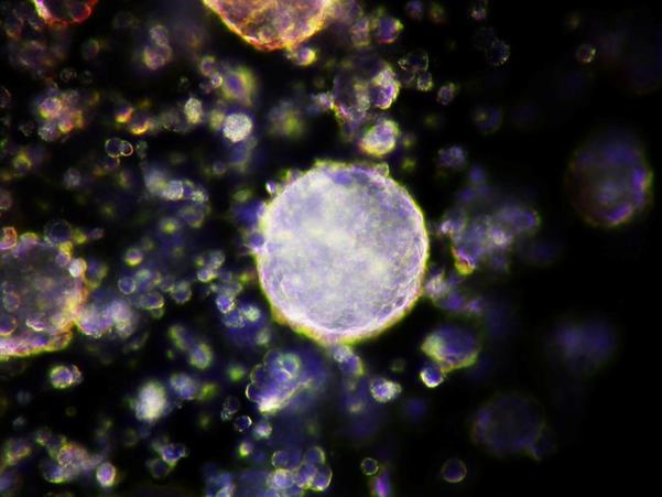
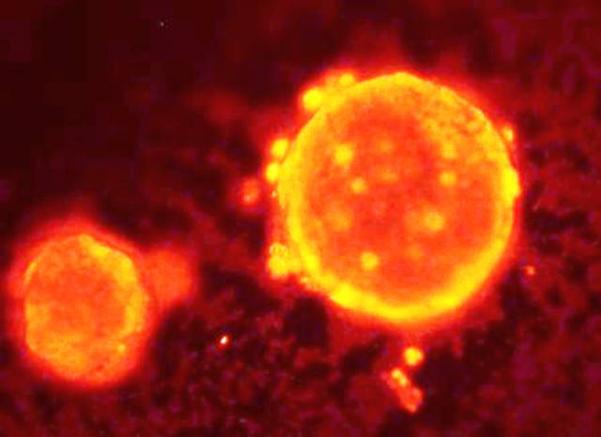
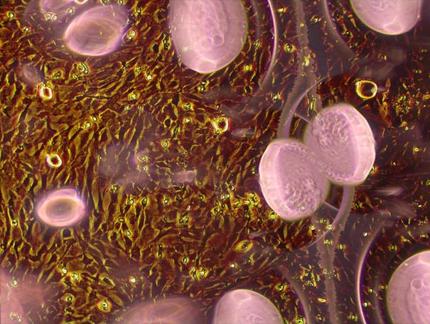
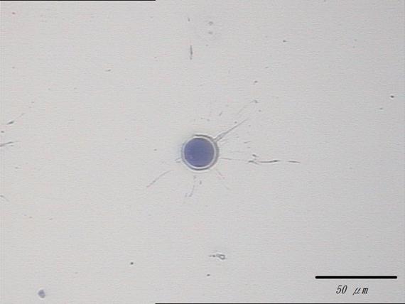
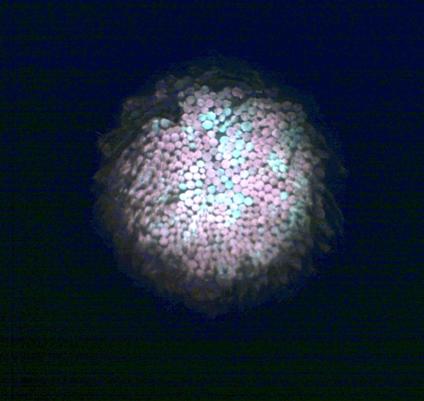
Title:
TINY BALL OF LIFE
Sneha Agarwal
School
of EPS
Microscopic view of an embryoid body. Dielectrophoresis is used to make
aggregates of mouse embryonic stem cells between electrode castellation. The
cells are arranged in a tightly packed 3D architecture to facilitate cell-cell
communication. Upon aggregation differentiation is initiated and the cells
begin to recapitulate embryonic development. Embryoid bodies are important as
they may serve as a good model to study earliest stages of development.
Title: TINY BALL OF LIFE
Sneha
Agarwal
School of EPS
Microscopic
view of an embryoid body. Dielectrophoresis is used to make aggregates of mouse
embryonic stem cells between electrode castellation. The cells are arranged in a
tightly packed 3D architecture to facilitate cell-cell communication. Upon
aggregation differentiation is initiated and the cells begin to recapitulate
embryonic development. Embryoid bodies are important as they may serve as a
good model to study earliest stages of development.
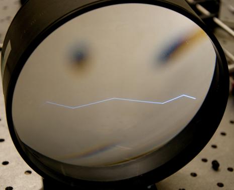
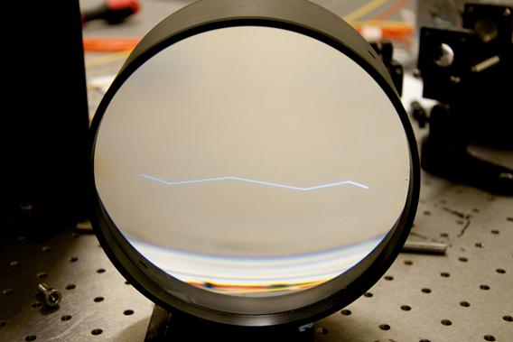
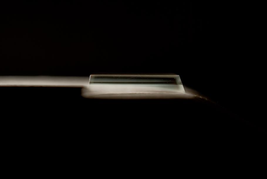
Title: Total internal reflection
Ryan
Warburton
School of EPS
The
photos help visualise total internal reflection; an important phenomenum which
demonstrates how optical fibres work.
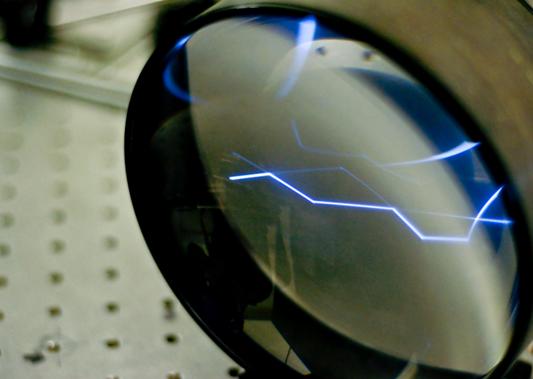
Title: Total internal reflection
Ryan
Warburton
School of EPS
The
photos help visualise total internal reflection; an important phenomenum which
demonstrates how optical fibres work.
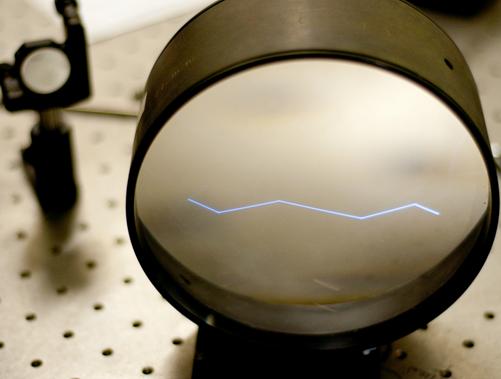
Title: Total internal reflection
Ryan
Warburton
School of EPS
The
photos help visualise total internal reflection; an important phenomenum which
demonstrates how optical fibres work.
Title: Total internal reflection
Ryan
Warburton
School of EPS
The
photos help visualise total internal reflection; an important phenomenum which
demonstrates how optical fibres work.
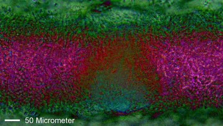
Title: Fibroblast
Rama
Yusvana
School of EPS
Fibroblast-type
morphology of embryonic skin cells
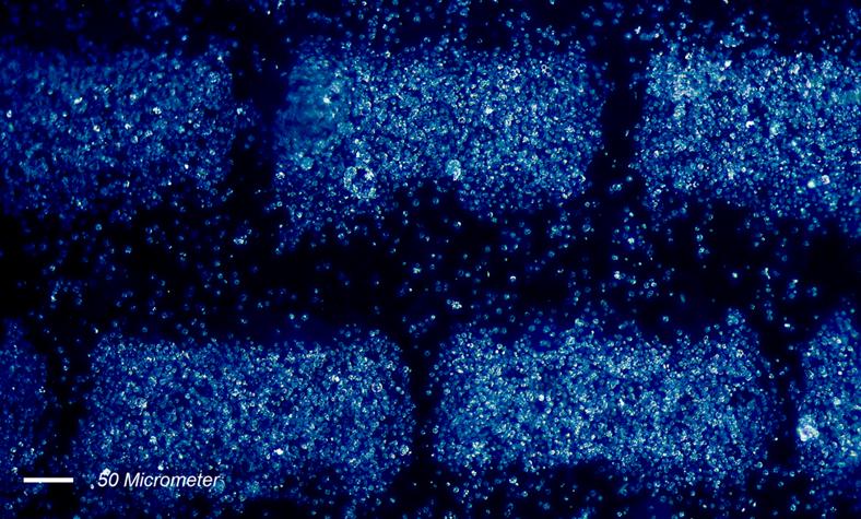
Title:
Cell Aggregates
Rama Yusvana
School
of EPS
Designing embryonic skin cell populations (dermal and epidermal) confined
in specific areas in defined geometrical pattern (Hexagonal), size and shape
between microelectrode array used for the study of developmental processes in
skin.
Title:
Line Aggregates2
Rama Yusvana
School
of EPS
Designing embryonic skin cell populations (dermal and epidermal) confined
in specific areas in defined geometrical pattern (Parallel Lines), size and shape
between microelectrode array used for the study of developmental processes in
skin.
Title:
Large Hexagonal
Rama Yusvana
School
of EPS
Designing embryonic skin cell populations (dermal and epidermal) confined
in specific areas in defined geometrical pattern (Hexagonal), size and shape
between microelectrode array used for the study of developmental processes in
skin.
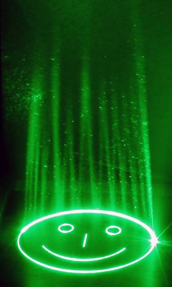
Title: Nanosecond Laser Marking
Rainer
Beck
School of EPS
Photograph
with long exposure time of laser marking of a metal plate using a nanosecond
laser machining workstation (wavelength 532nm) including a galvanometric scan
head. Visibility of laser beam enhanced by aerosol spray.
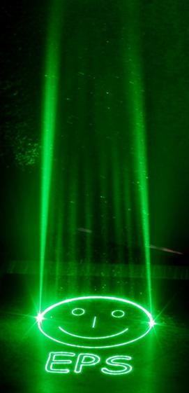
Title:
Nanosecond Laser Marking
Rainer Beck
School
of EPS
Photograph with long exposure time of laser marking of a metal plate
using a nanosecond laser machining workstation (wavelength 532nm) including a
galvanometric scan head. Visibility of laser beam enhanced by aerosol spray.
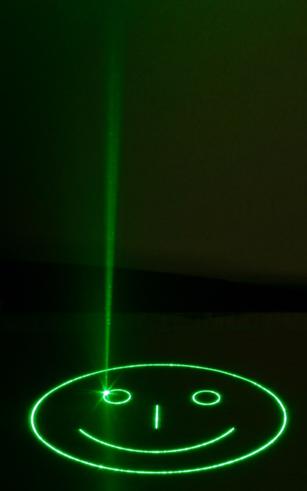
Title:
Nanosecond Laser Marking
Rainer Beck
School
of EPS
Photograph with long exposure time of laser marking of a metal plate
using a nanosecond laser machining workstation (wavelength 532nm) including a
galvanometric scan head. Visibility of laser beam enhanced by aerosol spray.
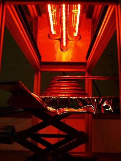
Title: solar heater powered by infrared
lights
Nikolaj
Rybakov
School of EPS
The
photo with spiral tube under red lights is a solar receiver. It is a model of
Donavan PHD student. The lights simulate the sun and for experimental reasons
it is tested with water flow through the receiver.
Title: -176ºC
Nikolaj
Rybakov
School of EPS
The
exposure control was really good. And with a hand in the photo, everything has
a life suddenly.
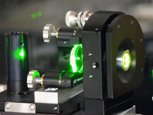
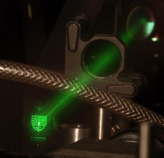
Title: A green laser beam going through
optics
Nikolaj
Rybakov
School of EPS
The
structure of the photo is clear and presents a serious "scientific"
mode. If it was taken on a tripod, that would be great.
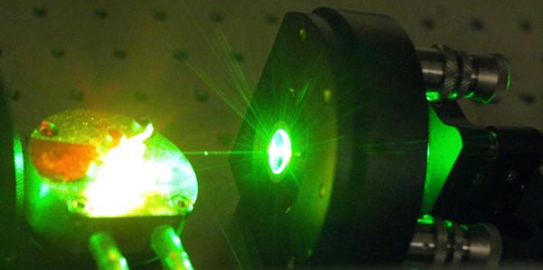
Title: Green laser
Nikolaj
Rybakov
School of EPS
A
green pumping laser beam focused on a Titanium doped sapphire crystal which is
the heart of a Kerr-lens mode-locking laser for generation of femtosecond
(10^-15 s) laser pulses. The latter is really good.
Title: Laser Drilling Glass
Wenlong
Chang
School of EPS
Glass
is a brittle material and it is hard to be manufactured by contact machining
process. The transparent glass is also hard to be machined by laser. This image
shows the drilling glass by laser machining. The good edge quality can be
achieved and obtained.
Wave Guide

Title: Concentrated Sunlight on Pipe
Steven Willis
The project
is to construct a Solar BBQ, where a copper pipe in the centre receives sunlight
reflected onto it using a reflective parabolic surface, this concentration
heats up the copper pipe, meaning it can cook a sausage! These pictures are of
the copper pipe heating up so much that the glue used to hold the insulation
has started to melt and smoke. 3972
is Colin Weighill writing down some results from the test.
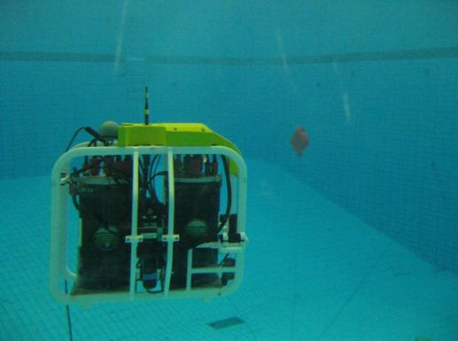
Title:
Looking for the target
Francesco Maurelli
At the Edinburgh Commonwealth
Pool, the AUV Nessie IV is looking for the target (orange buoy). Once detected,
it will follow it at a fixed distance
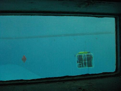
Title:
Target Acquired
Francesco Maurelli
At the Edinburgh Commonwealth Pool, the AUV Nessie IV has
acquired the target (orange buoy) and it is prepared to follow it at a fixed
distance.
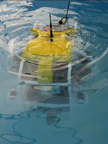
Title:
Nessie IV AUV
Francesco Maurelli
The autonomous underwater vehicle Nessie IV, developed by
the OSL/HWU. Winning entry for the second time in a row of the SAUC-E
competition.
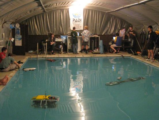
Title:
Hectic work at night
Francesco Maurelli
Somerton,
preparation for the SAUC-E Competition. Four teams are using the pool for basic
testing. The high temperature at day and the specific greenhouse environment
made the teams working mostly at night. The Heriot-Watt robot is with yellow
buoyancy on the bottom left and it will win the SAUC-E competition a few days
later this picture has been taken
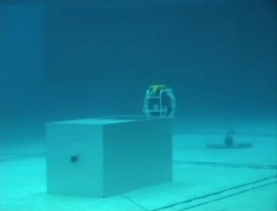
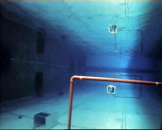
Title:
Autonomous Docking
Francesco Maurelli
SAUC-E Competition: HWU robot Nessie IV has autonomously
found the docking box and it is positioning to enter into it. It was the only
entrant able to successful perform this task. On the background, a ground
target is visible. The task was to find it and to hover at a specified
distance. Another task successfully completed by Nessie :-)
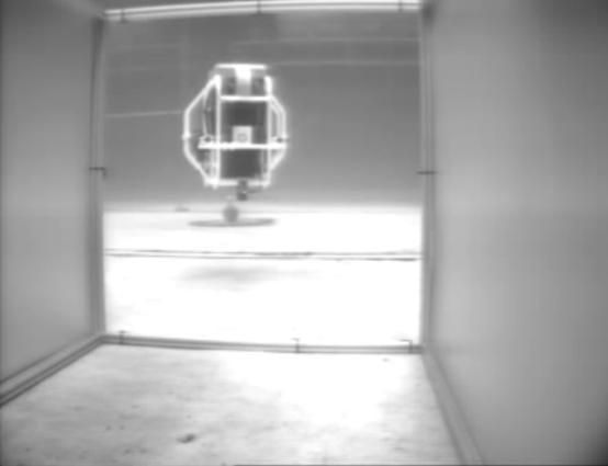
Title:
Underwater Competition
Francesco Maurelli
SAUC-E Competition: the underwater camera records the HWU
entry Nessie IV while it is passing through the gates. On the left handside, a
docking box is visible. A task of the competition was to autonomously find it
and to enter into it. Nessie IV was the only entry who managed to successfully
perform this task. Note the reflection on the surface. Nessie IV is very smart
with its camera not to get confused!
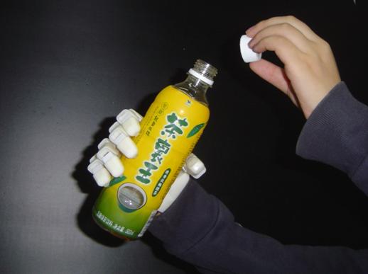
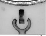
Title:
Under-Actuated Self-Adaptive Bionic Robotic Hand-Grasping Bottle
Guangbo
Hao
It has
potential applications as hand prosthesis for the person who has had an
amputation or as manipulator for grasping the complicated-shape objects
required in engineering surroundings. The under-actuated bionic robotic hand
has the advantages as follows. One
input to control multi-output motions; Simple control not using complicated
control system; Self-Adaptive grasping objectives; Possessing general hand
function; Low cost and light weight; Easy maintenance.
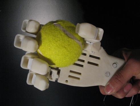
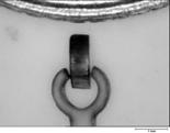
Under-Actuated
Self-Adaptive Bionic Robotic Hand--Grasping Ball
Guangbo
Hao
It has
potential applications as hand prosthesis for the person who has had an
amputation or as manipulator for grasping the complicated-shape objects
required in engineering surroundings.
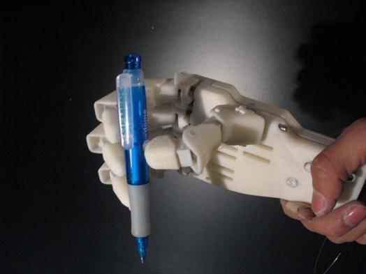
Under-Actuated
Self-Adaptive Bionic Robotic Hand--Grasping Pen
Guangbo
Hao
It has
potential applications as hand prosthesis for the person who has had an
amputation or as manipulator for grasping the complicated-shape objects
required in engineering surroundings.
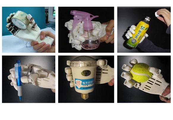
Under-Actuated
Self-Adaptive Bionic Robotic Hand-Grasping Different Objects
Guangbo
Hao
It has
potential applications as hand prosthesis for the person who has had an
amputation or as manipulator for grasping the complicated-shape objects
required in engineering surroundings.
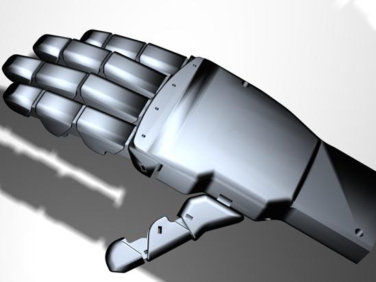
Under-Actuated
Self-Adaptive Bionic Robotic Hand-Simulation
Guangbo
Hao
It has
potential applications as hand prosthesis for the person who has had an
amputation or as manipulator for grasping the complicated-shape objects
required in engineering surroundings.
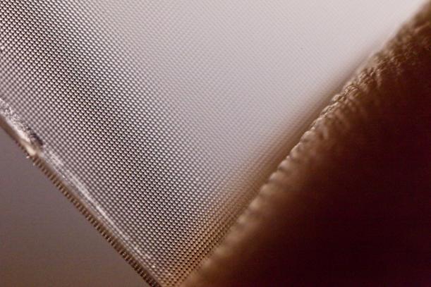
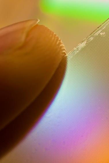
Title: Microlenses from a Plenoptic
camera
Thomas
Bishop
School of EPS
The
microlens array from a plenoptic camera, with a fingertip shown for scale. The lenses give repeated images inside a
plenoptic camera, allowing 3D imaging and synthetic refocusing to be applied in
software. Each microlens has a
135micron diameter and 0.5mm focal length.
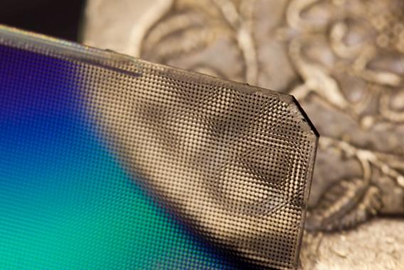
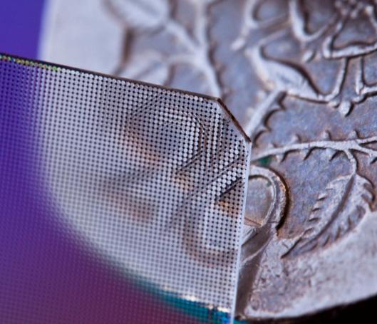
Title: Microlenses from a Plenoptic
camera
Thomas
Bishop
School of EPS
The
microlens array from a plenoptic camera, placed in front of a 20p coin showing
the effect of repetitions that the array gives inside a camera. The repeated images allow 3D imaging and
synthetic refocusing to be applied in software. Each microlens has a 135micron diameter
and 0.5mm focal length.
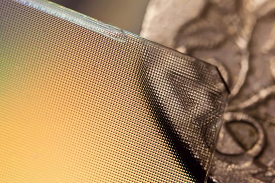
Title: Microlenses from a Plenoptic
camera
Thomas
Bishop
School of EPS
The
microlens array from a plenoptic camera is placed in front of a 20 pence coin to
show scale, and the effect of repetitions that the array gives inside a
camera. Each microlens is
135microns in diameter, with a 0.5mm focal length.
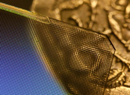
Title: Microlenses from a Plenoptic
camera
Thomas
Bishop
School of EPS
The
microlens array from a plenoptic camera is placed in front of a 20 pence coin
to show scale, and the effect of repetitions that the array gives inside a
camera. Each microlens is
135microns in diameter, with a 0.5mm focal length.
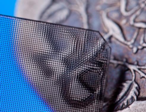
Title: Microlenses from a Plenoptic
camera
Thomas
Bishop
School of EPS
The
microlens array from a plenoptic camera, placed in front of a 20p coin showing
the effect of repetitions that the array gives inside a camera. The repeated images allow 3D imaging and
synthetic refocusing to be applied in software. Each microlens has a 135micron diameter
and 0.5mm focal length.
Title: Tissue Microlenses from a
Plenoptic camera
Thomas
Bishop
School of EPS
The
microlens array from a plenoptic camera, placed in front of a 20p coin showing
the effect of repetitions that the array gives inside a camera. The repeated images allow 3D imaging and
synthetic refocusing to be applied in software. Each microlens has a 135micron diameter
and 0.5mm focal length.
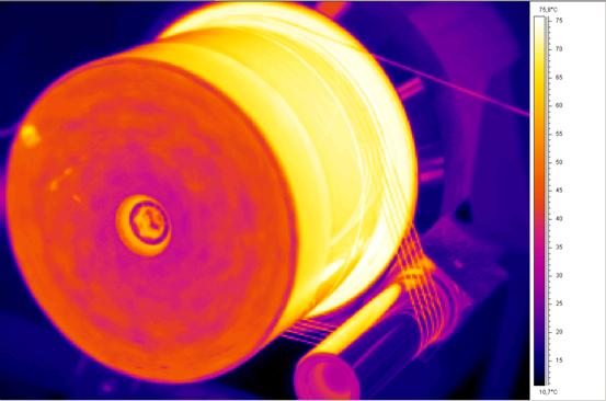
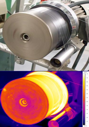
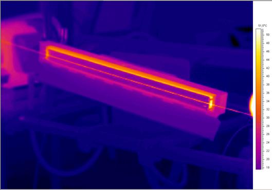
Title: Infrared Image of a Very Fine
Biodegradable Yarn in Hot Drawing Process
Basel Younes
A fine multi-filament yarn of 74 filaments, biodegradable aliphatic
aromatic co-polyester yarn, are drawn on hot drawing roller at 75 OC
in the hot drawing process, IR image illustrates the temperature variation to
get the temperature profile scale along or across the fibres and the roller, image colours correspond to the temperature scale on the
right.
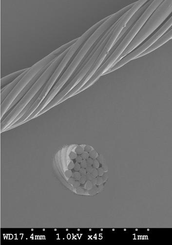
Title:
SEM Photo of Man Made Petroleum Based Biodegradable Yarn
Basel Younes
A clear photo of multi-filament yarn
of 30 filaments, biodegradable aliphatic aromatic co-polyester yarn, was taken
using a high magnification scanning electron microscope to show the surface and
the cross section of produced fibres in term of product quality investigation.
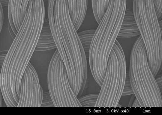
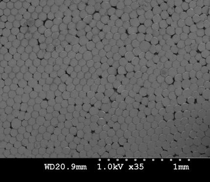
Title: SEM Photo of Biodegradable
Aliphatic Aromatic Co-Polyester Knitted Fabric
Basel Younes
A clear photo of knitted fabric made
of continuous multi-filament yarn, biodegradable aliphatic aromatic
co-polyester yarn, was taken using a high magnification scanning electron
microscope to show the surface of produced fabrics in term of product quality
and uniformity investigation.
Title: Super Optical Microscope Photo of the Cross Section of Biodegradable
Aliphatic Aromatic Co-Polyester Yarn
Basel Younes
An optical photo of cross section of
fine continuous multi-filament yarn, biodegradable aliphatic aromatic
co-polyeste
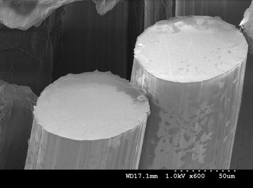
Title: Super SEM Photo of the Cross Section of Biodegradable Petroleum Based Fibres
Basel Younes
A clear photo of cross section of
biodegradable aliphatic aromatic co-polyester fibres was taken using a high
magnification scanning electron microscope to show the cross section variation
of produced fibres in term of product quality investigation.
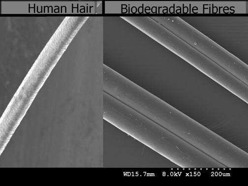
Title: Super SEM Photo to Compare
between Human Hair and the fine Produced Biodegradable Fibres
Basel Younes
A super photo of fine biodegradable
aliphatic aromatic co-polyester fibres was taken using a high magnification
scanning electron microscope to show the size of the fine produced fibres and
to compare it with human hair.
Title:
Super SEM Photo of Produced Biodegradable Fibres
Basel Younes
A super photo of fine biodegradable
aliphatic aromatic co-polyester fibres was taken using a high magnification
scanning electron microscope.
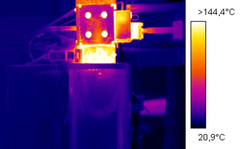
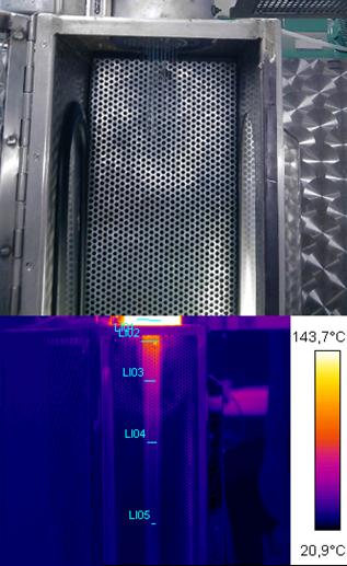
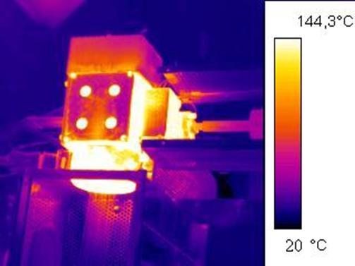
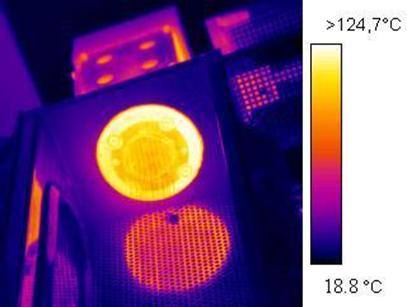
Title: Infrared Image of a Spinneret
and Die Head of Spinning Machine
Basel Younes
Infrared image of the spinneret and
die head of spinning machine illustrates the temperature variation to get the
temperature profile scale along or across the studied parts in term of heating
stability investigation, image colours correspond to the temperature scale on the
right.
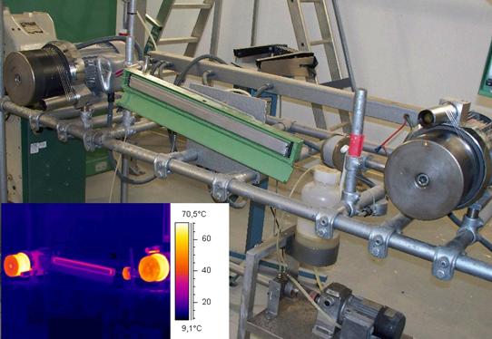
Title: From Optical Image to Infrared
Image of drawing of a very fine biodegradable yarn
Basel Younes
Depending on extensive thermal
analysis, a fine multi-filament yarn of 74 filaments, biodegradable aliphatic aromatic
co-polyester yarn, are drawn on hot drawing rollers at 60 OC in the hot drawing
process. With original view, IR image illustrates the temperature variation to
get the temperature profile scale along or across the fibres, image colours
correspond to the temperature scale on the right.
Title: From Optical Image to Infrared
Image of drawing of a very fine biodegradable yarn
Basel Younes
Depending on extensive thermal
analysis, a fine multi-filament yarn of 74 filaments, biodegradable aliphatic
aromatic co-polyester yarn, are drawn on hot drawing roller at 75 OC in the hot
drawing process. With original view, IR image illustrates the temperature
variation to get the temperature profile scale along or across the fibres,
image colours correspond to the temperature scale on the right.
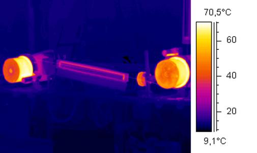
Title:
Infrared Image of drawing of a very fine biodegradable yarn
Basel Younes
Depending on extensive thermal
analysis, a fine multi-filament yarn of 74 filaments, biodegradable aliphatic aromatic
co-polyester yarn, are drawn on hot drawing rollers and plate at 60 OC in the
hot drawing process. IR image illustrates the temperature variation to get the
temperature profile scale along or across the fibres, image colours correspond
to the temperature scale on the right.
Title: From Optical Image to Infrared
Image of a very fine biodegradable spun yarn in cooling window of designed
experiment
Basel Younes
Depending on extensive thermal
analysis, a fine multi-filament yarn of 74 filaments, biodegradable aliphatic
aromatic co-polyester yarn, are spun at 140 OC in melt spinning process. With
original view, IR image illustrates the temperature variation to get the
temperature profile scale along or across the fibres in term of investigation
of cooling system quality, image colours correspond to the temperature scale on
the right.
Title: Infrared Image of a very fine
biodegradable yarn on controlled hot plate
Basel Younes
Depending on extensive thermal
analysis, a fine multi-filament yarn of 74 filaments, biodegradable aliphatic
aromatic co-polyester yarn, are drawn on hot plate at 50 OC in the hot drawing
process. IR image illustrates the temperature variation to get the temperature
profile scale along the fibres, image colours correspond to the temperature
scale on the right.
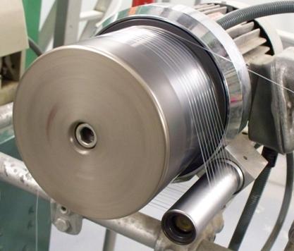
Title:
Optical Image of drawing of a very fine biodegradable yarn
Basel Younes
A fine multi-filament yarn of 74
filaments, biodegradable aliphatic aromatic co-polyester yarn, is drawn on hot drawing
roller at 75 OC in the hot drawing process.
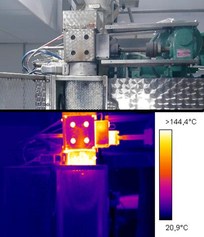
Title: Infrared Image of a Spinneret
and Die Head of Spinning Machine
Basel Younes
Infrared image of a spinneret and die
head of spinning machine illustrates the temperature variation to get the
temperature profile scale along or across the studied parts in term of heating
stability investigation, image colours correspond to the temperature scale on
the right.
Title: Infrared Image of a Spinneret
and Die Head of Spinning Machine
Basel Younes
Infrared image of the heat reflection
of a heated spinneret on the chassis, it illustrates the temperature variation
to get the temperature profile scale along or across the spinneret in term of
heating stability investigation, image colours correspond to the temperature
scale on the right.
Title: From Optical Image to Infrared
Image of drawing of a very fine biodegradable yarn
Basel Younes
Depending on extensive thermal
analysis, a fine multi-filament yarn of 74 filaments, biodegradable aliphatic
aromatic co-polyester yarn, are drawn on hot drawing rollers and plates at 60
OC in the hot drawing process. With original view, IR image illustrates the
temperature variation to get the temperature profile scale along or across the
process, image colours correspond to the temperature scale on the right.
Title: From Optical Image to Infrared
Image of the Die Head, Spinneret and Cooling Window of Melt Spinning Machine
Basel Younes
Depending on extensive thermal
analysis, die head, spinneret and cooling window of melt spinning machine at
140 oC process in melt spinning process. With original view, IR image
illustrates the temperature variation to get the temperature profile scale
along or across the cooling system in term of investigation of cooling system
quality, image colours correspond to the temperature scale on the right.